
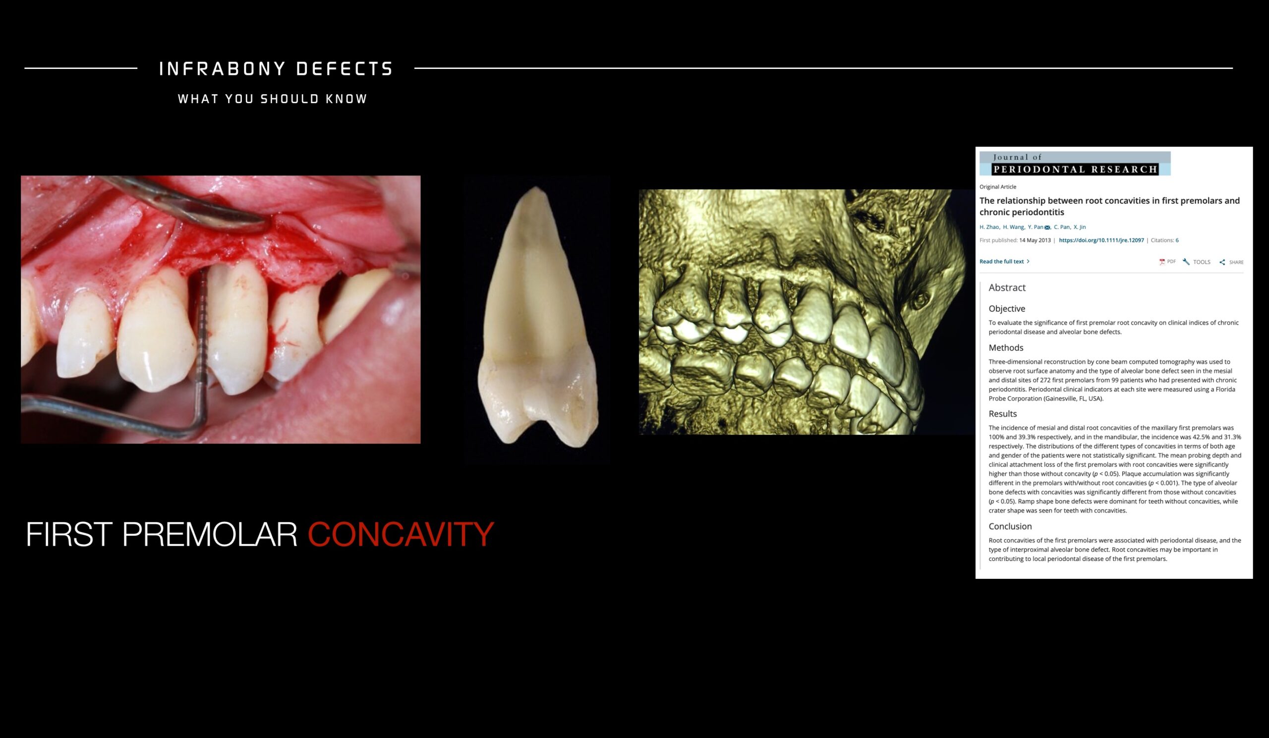
The root anatomy of the upper first premolars should be similar and at the same time different among individuals.
The anatomical reality is that on its formation at the most coronal aspect of the root a marked concavity at the mesial side is present, next to the canine creating a difficult architecture for proper periodontal tissue apposition, more prone residual food storage, difficult cleaning and proliferative bacterial growth.
This difficult architecture in addition to external factors that will increase the chances to pocket formation will make this area a high risk of non detected pockets which might evolve into infra bony defects and inter proximal alveolar bone defects establishing a higher risk of premolar and canine damage.
We usually never take this area as a risk in terms of potential development of local periodontal disease and we should probe first premolar areas even though it doesn’t present inflammation.
For the same reason that we focus in furcation involvement as risk areas especially in chronic periodontal disease patients we should take this areas seriously managing prosthetics, orthodontic movements that might trigger inter proximal alveolar bone loss becoming severe defects in the aesthetic zone (watch Severe Defect after Orthodontic treatment Masterclass) and recommend this patients keep the area away from plaque formation by flossing properly and treating this areas according to the protocols established to reduce pocket formation.