
For these reasons and more, you should visualize with CBCT certain aspects of the areas to treat.
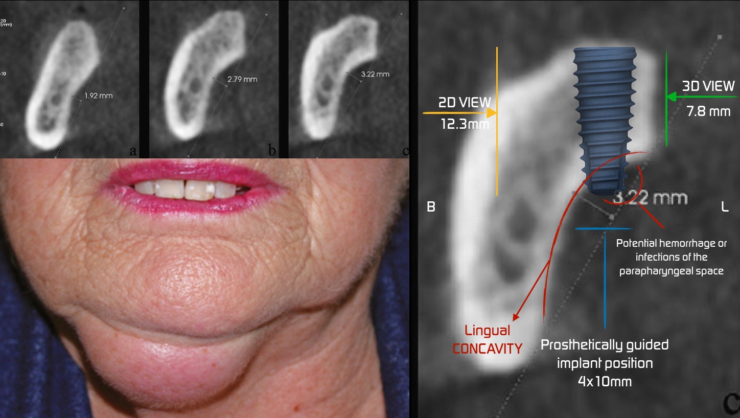
Lingual concavity or undercut is a common finding in the posterior mandible, and ignoring this undercut may result in perforation of the lingual cortex. (1)
Traumatisation of vital anatomical structures, neurologic injuries and severe bleeding in the floor of the mouth, which can be fatal if it causes upper airway obstruction traumatisation of vital anatomical structures, neurologic injuries and severe bleeding in the floor of the mouth, which can be fatal if it causes upper airway obstruction traumatisation of vital anatomical structures, neurologic injuries and severe bleeding in the floor of the mouth, which can be fatal if it causes upper airway obstruction (2)
Complications may occur during surgery, in the recovery phase or even after loading. In most cases, it is necessary to ensure accurate positioning of drill and implant fixture by clinical and radiographic examination of the areas where bone needs to be removed(3)
In order to prevent these complications, accurate radiographs are required to quantify the amount of bone, assess the location of adjacent anatomical structures and select the proper size of fixture and proper implant placement site. Radiography can be a valuable surgical guide for this purpose.(4)
Periapical and panoramic radiographs are often used to assess the site of implant placement. However, these techniques are two-dimensional and cannot provide adequate information about the bone width.(5)
Cross-sectional analysis using CBCT is suitable for assessment of lingual undercuts and prevention of lingual cortex perforation and subsequent complications. It has been proven that CBCT is beneficial when visualisation of a second plane is required prior to implant placement. (6 and 7)
Mandibular Lingual Concavity: A Crosssectional Analysis using Cone Beam Computed Tomography Fatemeh Salemi1 , Abbas Shokri2 , Maryam Foroozandeh3 , Manoochehr Karami4 , Zahra Khalili Journal of Clinical and Diagnostic Research, 2018, Oct, Vol-12(10): ZC37-ZC41
(1.)Chan HL, Benavides E, Yeh CY, Fu JH, Rudek IE, Wang HL. Risk assessment of lingual plate perforation in posterior mandibular region: a virtual implant placement study using cone-beam computed tomography. J Periodontol. 2011;82(1):129-35.Chan HL, Benavides E, Yeh CY, Fu JH, Rudek IE, Wang HL. Risk assessment of lingual plate perforation in posterior mandibular region: a virtual implant placement study using cone-beam computed tomography. J Periodontol. 2011;82(1):129-35.
(2.)Leong DJ, Chan HL, Yeh CY, Takarakis N, Fu JH, Wang HL. Risk of lingual plate perforation during implant placement in the posteriormandible: a human cadaver study. Implant Dentistry. 2011;20(5):360-63
(3.)Chan HL, Brooks SL, Fu JH, Yeh CY, Rudek I, Wang HL. Cross-sectional analysis of the mandibular lingual concavity using cone beam computed tomography. Clin Oral Implants Res. 2011;22(2):201-06.
(4.)Quirynen M, Mraiwa N, van Steenberghe D, Jacobs R. Morphology and dimensions of the mandibular jaw bone in the interforaminal region in patients requiring implants in the distal areas. Clin Oral Implants Res. 2003;14(3):280-85
(5.)Chen LC, Lundgren T, Hallstrom H, Cherel F. Comparison of different methods of assessing alveolar ridge dimensions prior to dental implant placement. J Periodontol. 2008;79(3):401-05.
(6.)Nickenig HJ, Wichmann M, Eitner S, Zoller JE, Kreppel M. Lingual concavities in the mandible: a morphological study using cross-sectional analysis determined by CBCT. J Craniomaxillofac Surg. 2015;43(2):254-59.
(7.)Kaeppler G, Mast M. Indications for cone-beam computed tomography in the area of oral and maxillofacial surgery. Int J Comput Dent. 2012;15(4):271-86.