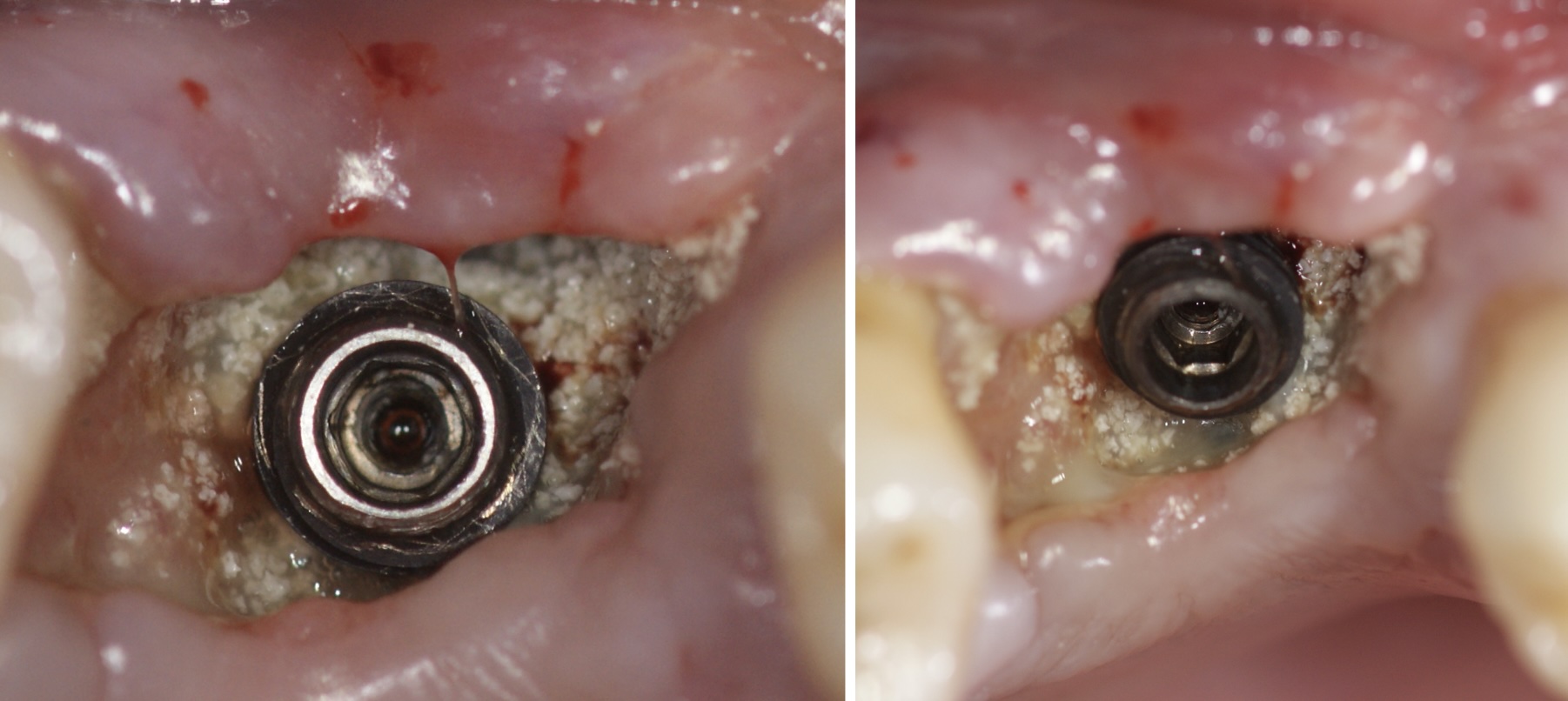
Peri-implantitis is one of the most challenging complications in modern implantology. Despite technological advancements and surgical techniques, inflammation and bone loss around implants remain frequent issues that threaten treatment longevity. For this reason, early diagnosis of peri-implantitis is essential to intervene promptly and prevent implant failure.
This article provides a comprehensive overview of the signs, diagnostic techniques, and tools to identify peri-implantitis in its early stages, offering a complete guide for you, the dental professional.
What Is Peri-Implantitis and How Does It Develop?
Peri-implantitis is an inflammatory process affecting the peri-implant tissues, leading to progressive bone loss around the implant. Its progression can be divided into three stages:
- Initial Stage: Peri-Implant Mucositis
- Reversible inflammation of the soft tissues surrounding the implant.
- Mainly caused by biofilm accumulation and poor oral hygiene.
- No detectable bone loss is observed.
- Intermediate Stage: Early Peri-Implantitis
- Initial bone resorption around the implant begins.
- Symptoms are mild, making diagnosis challenging without specific tools.
- Advanced Stage: Severe Peri-Implantitis
- Significant bone loss occurs.
- In severe cases, implant mobility may be observed.
Risk Factors Associated with Peri-Implantitis
Identifying patients at higher risk is crucial to prevent peri-implantitis. Common risk factors include:
- History of Periodontal Disease: Patients with a history of periodontitis are more susceptible.
- Poor Oral Hygiene: Biofilm accumulation is the primary trigger.
- Smoking: Impairs healing capacity and increases inflammation.
- Uncontrolled Diabetes: Affects the host’s immune response.
- Implant and Prosthetic Design: Rough surfaces or unfavorable connections can promote bacterial colonization.
- Biomechanical Overload: Improper occlusion can lead to microfractures in peri-implant bone.
Clinical and Radiographic Signs of Peri-Implantitis
Early diagnosis requires a thorough analysis of the following signs:
Clinical Signs
- Soft Tissue Inflammation: Redness, swelling, and bleeding upon probing.
- Increased Probing Depths: Peri-implant pockets greater than 4-5 mm.
- Suppuration: Purulent discharge when applying pressure to the peri-implant mucosa.
- Pain: Rare but may be reported by some patients.
Radiographic Signs
- Marginal Bone Loss: Bone loss around the implant threads.
- Changes in Bone Density: Radiolucent areas surrounding the implant.
- Compare with baseline radiographs to assess progression.
Tools for Early Diagnosis
Advanced technologies complement clinical examinations:
1. High-Resolution Periapical Radiographs
- Detect early bone changes.
2. Peri-Implant Probes
- Plastic or titanium probes prevent implant surface damage.
- Measure probing depth and bleeding on probing.
3. Microbiological Analysis
- Identifies specific pathogens associated with peri-implantitis, such as Porphyromonas gingivalis.
4. Laser Fluorescence (PerioScan)
- Detects residual biofilm on peri-implant surfaces.
5. Cone-Beam Computed Tomography (CBCT)
- Provides 3D images to evaluate bone loss in detail.
Monitoring Protocols for Early Diagnosis
- Regular Follow-Ups
- Schedule check-ups every 3-6 months for at-risk patients.
- Perform comprehensive periodontal records at each visit.
- Establish Baseline Data
- Document radiographs and probing depths at implant placement.
- Patient Education
- Provide clear instructions on oral hygiene and maintenance.
What to Do After an Early Diagnosis?
Once peri-implantitis is detected in its early stages, immediate intervention is critical:
- Implant Surface Decontamination
- Use ultrasonic scalers, titanium curettes, or low-power lasers.
- Infection Control
- Local or systemic antibiotic therapy depending on the severity.
- Guided Bone Regeneration (GBR)
- Use biomaterials and membranes to treat significant bone defects.
- Risk Factor Management
- Modify patient habits, such as quitting smoking or improving glycemic control as well as Vitamin D levels.
Conclusion
The long-term success of implants depends not only on precise surgical placement but also on rigorous follow-up and early diagnosis of complications like peri-implantitis. As dental professionals, it is our responsibility to implement advanced prevention and diagnostic protocols to ensure peri-implant health for our patients.
Your Practice, Your Responsibility
Proactive management is always more effective than reactive treatment. Incorporate these strategies into your daily practice and contribute to raising the standards of modern implantology.

